CN

RetiView 500

Optical coherence tomography (OCT)

OCT RetiView 500 can perform tomographic imaging on the macular area, optic disc, choroid and other tissues of the fundus, providing important diagnostic basis and treatment for eye diseases, especially fundus diseases, glaucoma screening, diagnosis, follow-up and efficacy evaluation, a high-resolution, non-contact, non-invasive imaging technique for easy and fast examination.

01

Fully automatic

Fully automatic one-key operation, automatic eye tracking, automatic tomography of the macula, optic disc and other parts

Capture images automatically

02

Comprehensive measurement analysis tools

• Up to 14 types of analysis, accurate and comprehensive analysis of the fundus
• The use of OCT to measure macular hole size before eye surgery allows doctors to objectively assess the effect of macular hole surgery, and the changes in diabetic macular edema can be accurately and predictably measured by OCT
• Possibility to diagnose fine vitreomacular traction and provide accurate pre- and post-operative macular thickness assessment
• Can accurately measure subtle changes in retinal structure and make precise analysis of the retinal nerve fiber layer


Provide more detailed analysis data through precise analysis of ganglion cell layer-inner plexiform layer

Glaucoma analysis function, providing more accurate optic disc area, cup-to-disk ratio and other data services

Radial analysis of the macula for richer and more accurate data

03

Rich image editing software


Fundus OCT synthetic enface, automatic brightness and contrast adjustment

Macular 3D data acquisition, automatic segmentation of 7-layer boundaries after saving, and entering into analysis to quickly generate a macular thickness topographic map

1-line high-resolution acquisition at any angle and in any area, instantly synthesizing high-definition results

5-line fast high-definition screening and collection in any area

04

Case Reports

Monocular comprehensive analysis, optic disc and RNFL analysis

Technical parameter

OCT
Methodology Spectral domain OCT
Axial resolution ≦6um(in tissue)
Transverse resolution ≦20(in tissue)
Scan depth ≥2.5mm(in air)
Scan range 6-12mm
Scan speed ≥24,000A-scan/sec,up to 36,000A-scan/sec
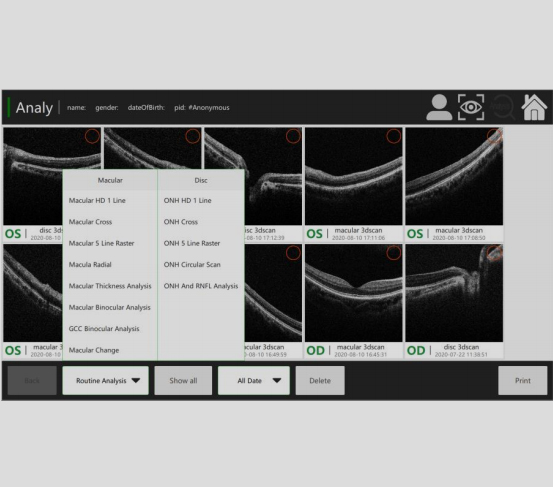
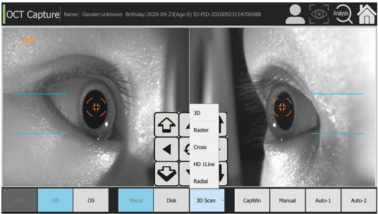
Scan mode 3D(Macular&Optic Disk) ,HD,Raster,Circle,Cross
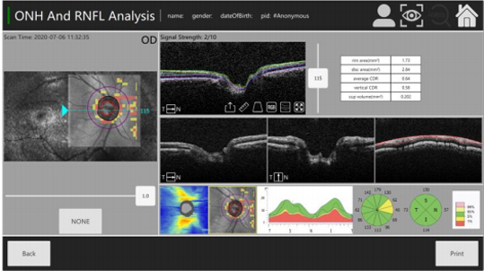
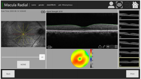
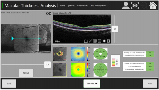
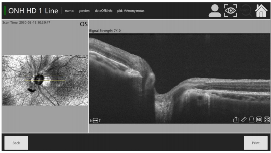
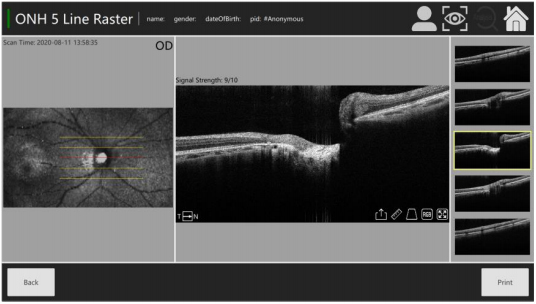
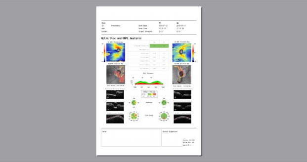
Analysis mode Up to 7 retinal layers segmentation,Macular analysis mode,RNFL&Optic Disk analysis mode,Glaucoma analysis mode and Progress analysis for follow-up examination.
Fundus image OCT en face
Focus adjustment -15D to+15D
Pupil diameter ≥3mm
OCT light source 840nm SLD
Operation 13.3inch touchscreen,optional external mouse or keyboard
Power supply 100-240V,50/60HZ
Dimensions 497x395x490mm
Weight 34kg